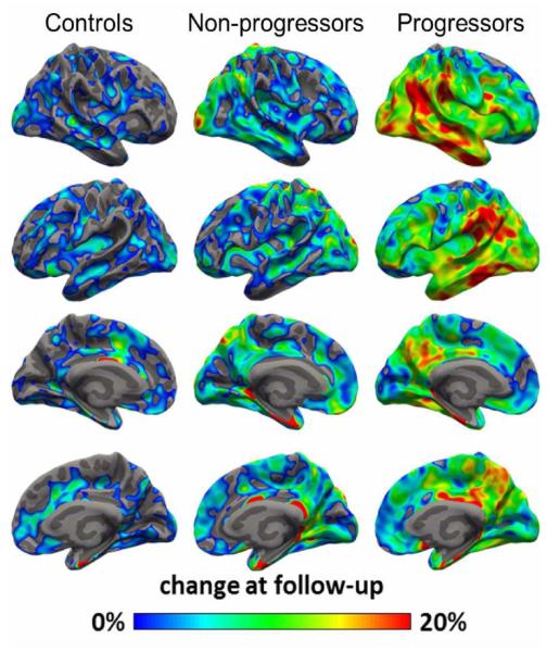
Fig 4.
Average percent change in 11C-PBR28 binding from baseline, overlaid on semi inflated cortical surface. Patients who showed clinical progression during the study interval (n = 9) had greater increases in 11C-PBR28 binding than non-progressors (n = 5) or controls (n = 8), with the greatest change observed in inferior temporal and parietal cortices.

