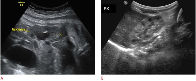
Fig. 2. Representative sonogram of transient hydronephrosis in a neonate.
Figure captionA. Isolated dilation of the renal pelvis is seen on prenatal ultrasound scan at 37 weeks of gestation. B. First postnatal ultrasonography, obtained at day 4 after birth, shows complete resolution of the dilation. RK, right kidney.

