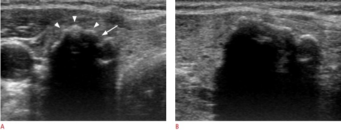
Fig. 1. Benign nodule with isolated macrocalcification in a 56-year-old woman.
Transverse sonogram (A) shows a calcified nodule (18 mm) with a lobulated contour (arrowheads) and interruption (arrow) of the anterior margin. The posterior margin of the calcified nodule is not visualized by strong posterior acoustic shadowing on transverse or longitudinal ultrasonography (A, B).

