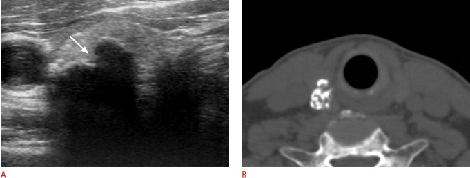
Fig. 4. Papillary carcinoma manifested as an isolated macrocalcification in a 62-year-old man.
A. Transverse sonogram shows a calcified nodule (16 mm) with lobulated contour and focal interruption (arrow) of the anterior margin. B. A computed tomography image demonstrates that the isolated macrocalcification correlates with conglomerated coarse calcifications. Suspicious metastatic lymph nodes were detected in the ipsilateral lateral and central neck by ultrasonography (not shown), and metastatic lymph nodes and extrathyroidal extension of the tumor were found by surgery.

