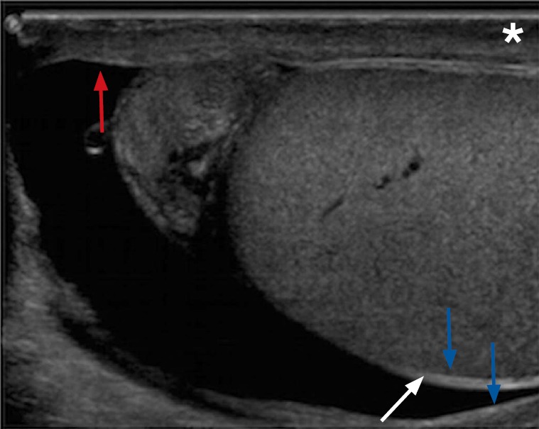
Fig. 1. Tunica albuginea and tunica vaginalis of a 22-year-old male with right scrotal pain.

Sonogram of the testicle effectively demonstrates the tunica albuginea which envelopes the testicle (red arrow). The testicle with its tunica albuginea is covered by the visceral layer of the tunica vaginalis (white arrow). The inner aspect of the scrotal wall (asterisk) is covered by the parietal layer of the tunica vaginalis (blue arrows). Normally both layers of the tunica vaginalis are only separated by a small amount of fluid; however, in this case there is a moderate amount of fluid separating the two layers that allows a good demonstration of the anatomy.
