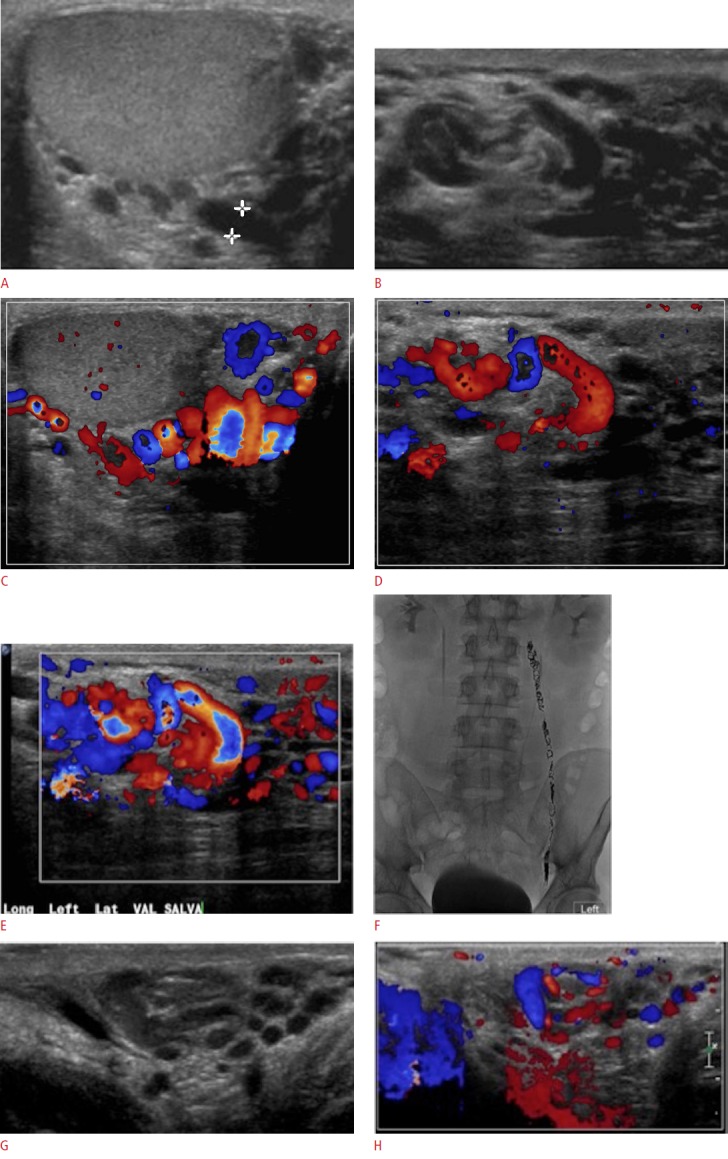
Fig. 15. Varicocele in a 33-year-old male with scrotal swelling.

A-D. Ultrasonogram reveals several tubular structures greater than 3 mm (A, B) with markedly increased vascular flow on color Doppler evaluation on resting (C, D). E. Increased vascular flow with engorgement of the veins is seen during Valsalva maneuver. F. The patient underwent left gonadal vein embolization with multiple endovascular coils seen in the region of the left gonadal vein on a subsequent abdominal radiograph. G, H. Follow-up ultrasonography demonstrates reduction in size of the varicoceles (G) with somewhat decreased vascular flow (H).
