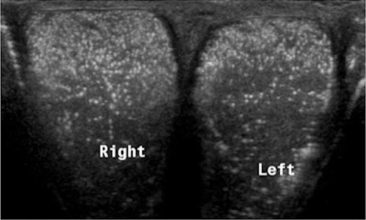
Fig. 17. Testicular microcalcifications in a 35-year-old patient with scrotal discomfort.

Comparison view of both testicles demonstrates multiple small echogenic foci within the testicular parenchyma. No underlying mass was identified.

Comparison view of both testicles demonstrates multiple small echogenic foci within the testicular parenchyma. No underlying mass was identified.