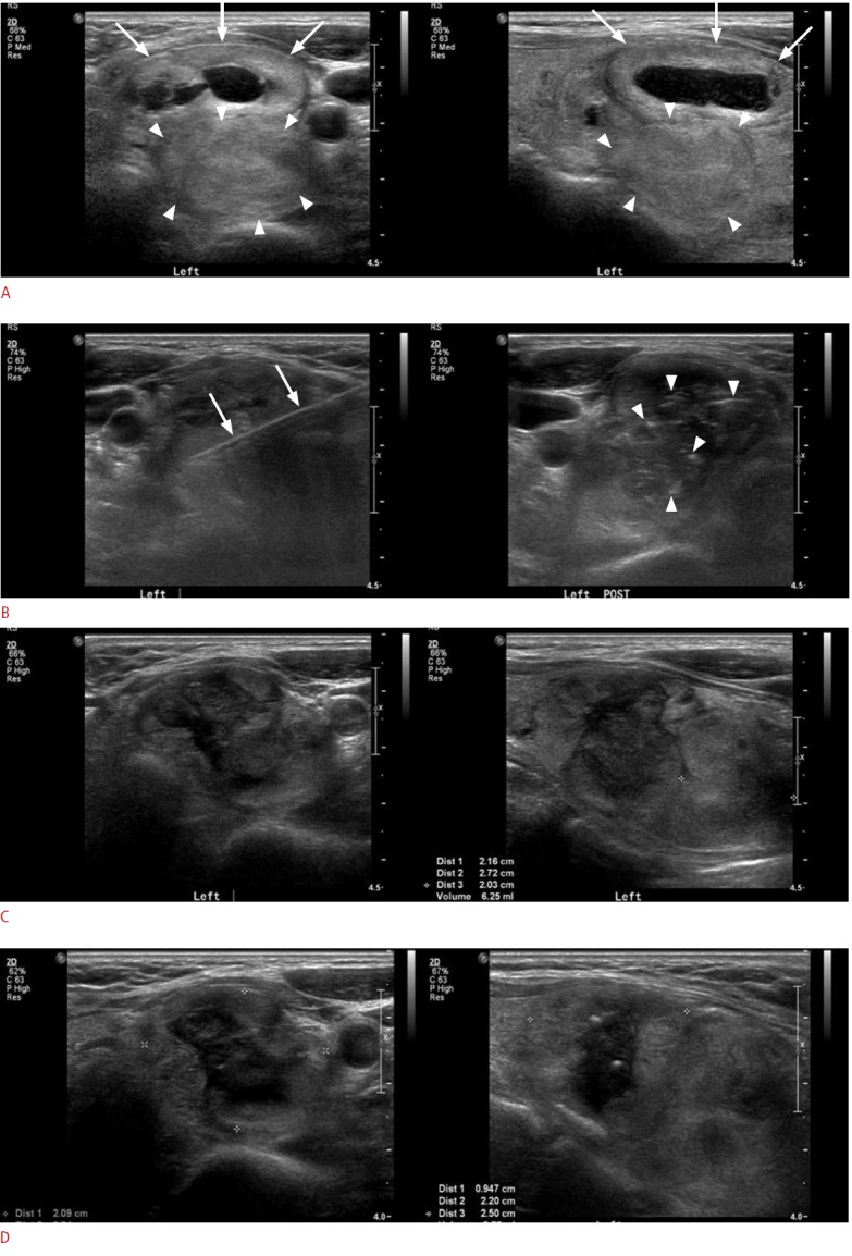
Fig. 2. A 60-year-old woman with two conglomerated solid nodules in the left thyroid.

A. Initial ultrasonogram shows a predominantly solid nodule (arrows) in the superficial area and a pure solid nodule (arrowheads) in the deep portion of the left thyroid abutting each other, with the deep nodule showing a relatively ill-defined margin in the lateral and posterior aspects in comparison with the superficial nodule. B. During the radiofrequency ablation, an internally cooled electrode (arrows) is inserted into the nodules and multiple echogenic micro-bubbles (arrowheads) were noted within the nodules after the procedure. C. On 1-month follow-up ultrasonography after the ablation, the nodules are slightly decreased in volume and size, with the volume reduction ratio of the superficial nodule being 36% and that of the deep nodule being 23%; thus a second session of radiofrequency ablation was performed for these nodules. D. On 1-year follow-up ultrasonography after the ablation, superficial and deep nodules show a greater decrease in volume (43% and 25%, respectively), but they do not achieve a volume reduction ratio of more than 50%; thus the therapeutic success criteria are not satisfied in this patient.
