
Fig. 2.
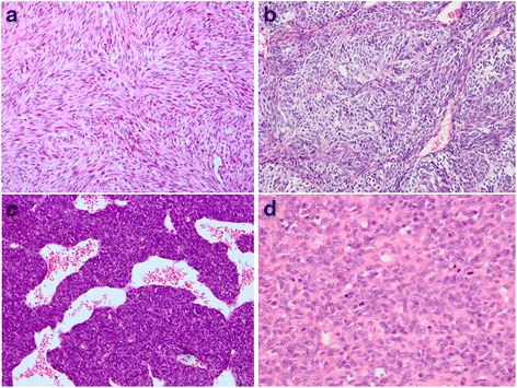
Microscopic features. a Monophasic synovial sarcoma, tumor consisting of uniform hyperchromatic spindled cells arranged in a prominent fascicular pattern (H&E, ×200). b Biphasic synovial sarcoma, composed of oval epithelial nests and surrounded by spindle cell components (H&E, ×200). c Poorly differentiated synovial sarcoma, showing high-grade spindle-shaped cells and a hemangiopericytoma-like vascular pattern, reminiscent of a malignant peripheral nerve sheath tumor (H&E, ×200). d Poorly differentiated synovial sarcoma, consisting of large epithelioid cells, indistinguishable from a poorly differentiated carcinoma or mesothelioma (H&E, ×400)