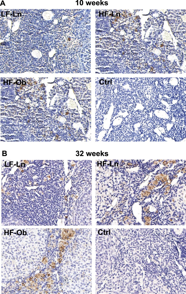
FIG. 4.
Representative images showing the presence of macrophages in the ovary. Macrophages were identified using marker CD68. Increased expression of macrophage marker CD68 was noted in both the HF-Ln (n = 6) and HF-Ob (n = 6) groups compared to the LF-Ln mice (n = 6), after 10 (A) and 32 (B) wk on HFD. These findings indicate increased tissue inflammation in the ovary after prolonged exposure to HFD regardless of obese phenotype. Ctrl, control. Original magnification ×200.

