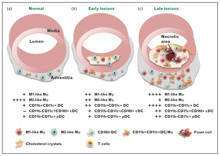
FIGURE 1.
Myeloid cells in atherosclerosis development. (a) The normal aorta contains some T lymphocytes and a small number of myeloid cells that express M2 markers and may be resident vascular macrophages. (b) Early atherosclerotic lesions are characterized by accumulation of myeloid cells in the subendothelial space. The number of cells in the adventitia is significantly increased. The macrophage population now represents a mixture of ‘M1-like’ and ‘M2-like’ macrophages. Various types of dendritic cells (DC) start to accumulate in both adventitia and plaque. (c) Late atherosclerotic lesions are characterized by accumulation of cholesterol crystals and various types of immune cells including foam cells, whose death leads to formation of the necrotic core, which is related to plaque instability and rupture. The macrophage population is mainly ‘M1-like’. High numbers of dendritic cell with potential pro-inflammatory and anti-inflammatory functions are detected in the adventitia and atherosclerotic plaque. High numbers of interactions between antigen-specific T cells and antigen-presenting cells (dendritic cell and macrophages) are observed in plaque and adventitia. pDC, plasmacytoid dendritic cell, cDC, conventional dendritic cell, Mϕ, macrophage.

