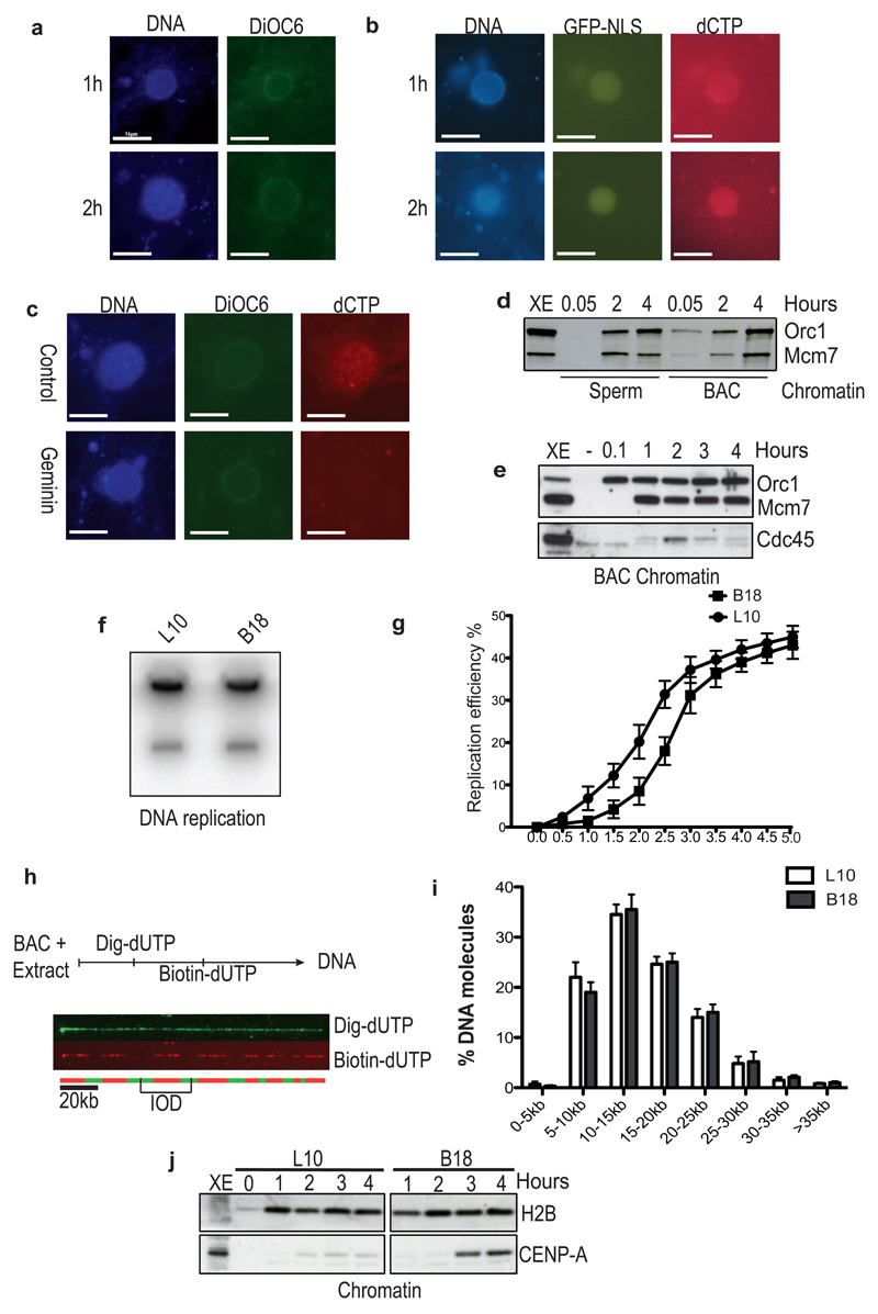
Figure 1. BAC DNA induced nuclei formation and DNA synthesis in interphase Xenopus egg extract.
(a) BACs were incubated in interphase extract for the indicated time. Samples were fixed and stained with DAPI for DNA (DNA) and DiOC6 for membranes (DiOC6). (b) Nuclei assembled in interphase extract supplemented with GFP-NLS and Cy3-dCTP (dCTP). (c) BACs replicated for 4 hours in egg extract supplemented with buffer (Control) or recombinant geminin (Geminin). (d) and (e) Chromatin isolated from sperm and BACs nuclei at different times and analysed by WB with the indicated antibodies. Representative images of experiments performed at least three times are shown. (f) Autoradiography of non-centromeric (L10) and centromeric (B18) BACs replicated in the presence of 32PdCTP. A representative image is shown. (g) Replication kinetics of non-centromeric L10 and centromeric B18 DNA. Error bars represent ± sd of the mean. n=3 experiments; p<0.001 when comparing L10 and B18 mean values for all the indicated times; unpaired two-tailed t-test. (h) Scheme of DNA combing experiment and example of DNA fiber visualization by immunofluorescence. BACs were incubated in egg extract, supplemented with Dig-dUTP (Green) and, at later time points, with Biotin-dUTP (Red). DNA was isolated at 3.5 hours from addition to egg extract for combing. Typical combing of L10 BAC is shown. Midpoints of green tracts (Dig-dUTP) represent origins of DNA replication (replication eye in the red track). Distance between midpoints of two adjacent replication eyes represents inter-origins distance (IOD) (i) Graph showing distribution percentage of IODs measured for control L10 (black) and B18 DNA (grey). At least hundred fibres were scored for each sample. Error bars represent ± sd of the mean. n=3 experiments; p<0.001 when comparing L10 and B18 mean values for all IODs; One-way Anova. (j) Chromatin was isolated at the indicated times of the replication reactions with L10 and B18 DNA and then analyzed by WB using the indicated antibodies. A representative of 3 experiments is shown. Uncropped gel images for all experiments are shown in Supplementary Figure 6.

