
Figure 2.
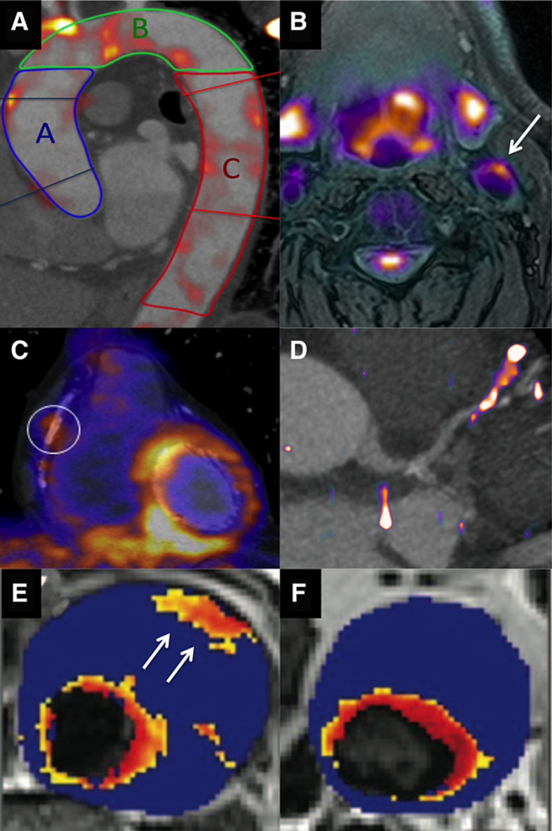
Molecular imaging of vascular inflammation activity. A, 18F-fluorodeoxyglucose (18F-FDG) positron emission tomography (PET) image fused with contrast computed tomographic (CT) angiogram of the thoracic aorta, demonstrating regions of increased activity in the ascending aorta (blue), aortic arch (green), and descending aorta (red). B, 18F-FDG PET/magnetic resonance (MR) image of the carotid arteries demonstrating increased activity in the left carotid artery and the excellent soft tissue contrast provided by MR. C, 18-FDG PET fused with a CT coronary angiogram demonstrating increased uptake in the left ventricle but also in a remote plaque in the mid right coronary artery. Image in panel C reproduced from Cheng et al21 with permission of the publisher. Copyright © 2012, the Society of Nuclear Medicine and Molecular Imaging, Inc. D, 68Ga-DOTATATE PET/CT image with increased activity localizing to a plaque in the mid left anterior descending artery. E, T2* Map from patients with an abdominal aortic aneurysm that had been administered ultra small particles of iron oxide (USPIO). A hot spot is observed in the anterior wall of the aneurysm (arrow). F, In a second patient, focal areas of increased USPIO uptake can be observed (the increased signal adjacent to the lumen is considered normal because of high signal in the blood pool). Images in panels E and F reproduced from Richards et al23 with permission of the publisher. Copyright © 2011, Wolters Kluwer Health, Inc.