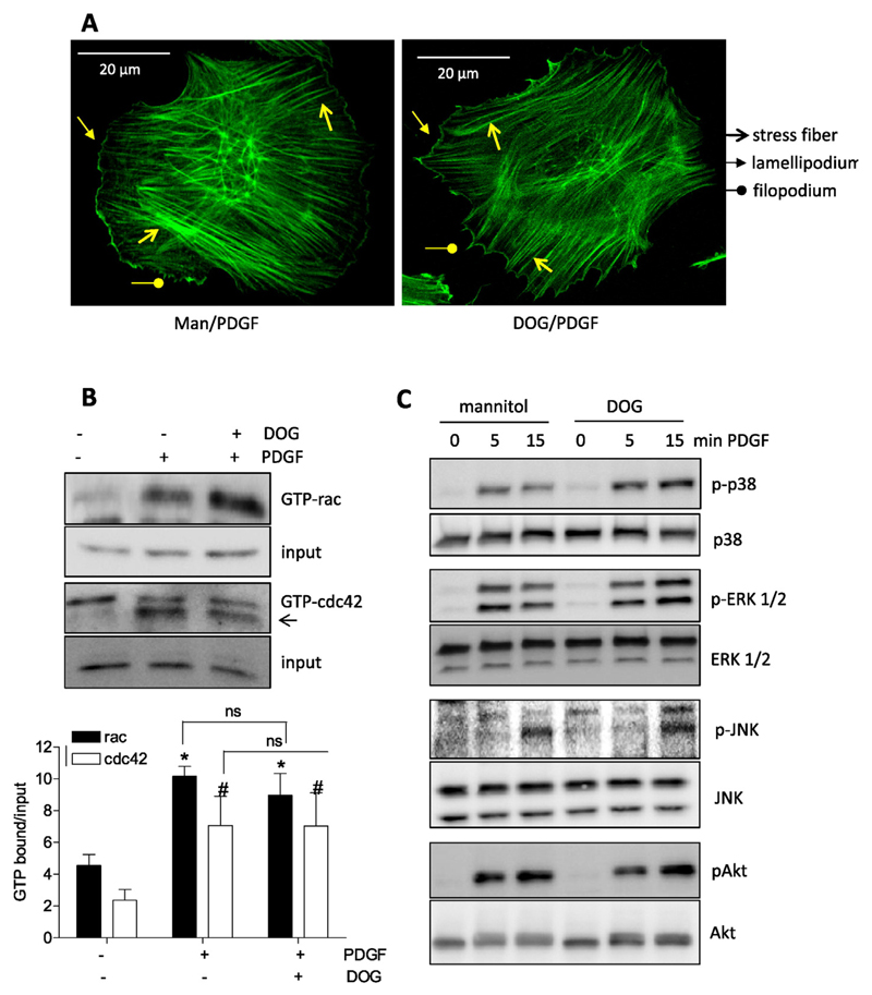
Fig. 3.
Inhibition of glycolysis does not interfere with early actin cytoskeleton reorganization. (A) Quiescent VSMC were stimulated with PDGF (10 ng/mL; 1 h) after 30 min pretreatment with mannitol (Man, 30 mM, osmotic control) or deoxyglucose (DOG, 30 mM). Cells were fixed, stained with FITC phalloidin and viewed under a confocal microscope. Stress fibers, lamelli –and filopodia are indicated with the appropriate arrows. (B) Quiescent VSMC were treated with 30 mM mannitol (−) or deoxyglucose (30 mM DOG) for 30 min as indicated before they were stimulated with PDGF (10 ng/mL) for 3 min. Lysates were then subjected to pulldown of GTP bound ras and cdc42. An aliquot served as input control. Representative immunoblots from three independent experiments ae depicted. The bar graph shows compiled densitometric data from all performed experiments. (n = 3, mean + SD.; *, # p < 0.05; ANOVA, Dunnett (vs respective unstimulated control). (C) Quiescent VSMC were treated with 30 mM mannitol (−) or deoxyglucose (30 mM DOG) for 30 min as indicated before they were stimulated with PDGF (10 ng/mL) for 5 or 15 min. Lysates were then subjected to immunoblots analyses as indicated. Representative blots from three independent experiments are depicted.

