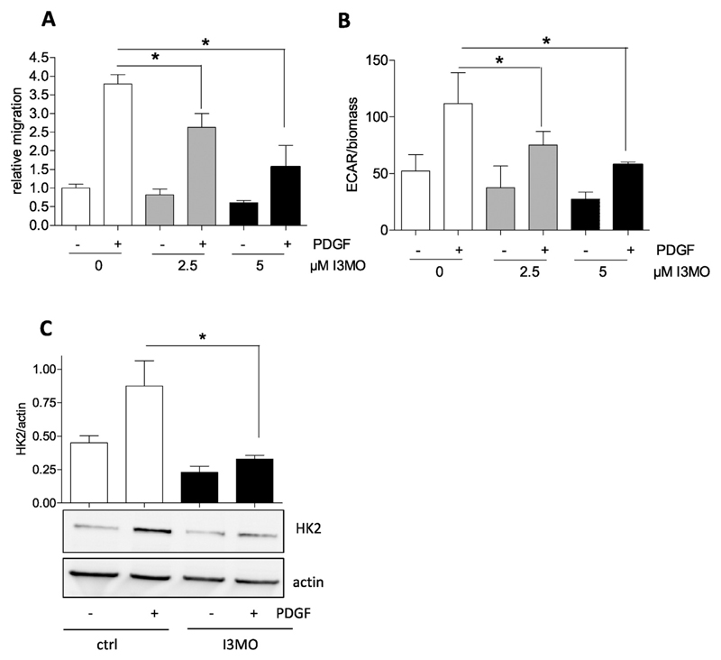
Fig. 4.
The antimigratory activity of I3MO is accompanied by a reduced glycolytic rate and diminished HK2 induction in PDGF-activated VSMC. (A) Serum-deprived VSMCs were subjected to a wound healing (scratch) assay with or without stimulation with 10 ng/ml PDGF-BB in the presence of 2.5 and 5 μM I3MO. Graphs indicate relative values of wound closure as assessed by CellProfiler Software (n = 3, mean + SD.; * p < 0.05; Student’s t-test). (B) Glycolytic activity in quiescent and PDGF-stimulated (6 h) VSMC in the absence or presence of I3MO (2.5 and 5 μM) was determined by extracellular flux analysis as described. The bar graph depicts compiled data of three independent experiments (n = 3, mean + SD.; * p < 0.05; Student’s t-test). (C) Serum-deprived VSMC were pretreated with 5 μM I3MO and then stimulated with PDGF (10 ng/mL) for 6 h. Total cell lysates were subjected to immunoblot analysis for HK2 and actin. Representative blots from three independent experiments and compiled densitometric analysis are depicted (n = 3, mean + SD.; * p < 0.05; ANOVA, Bonferroni).

