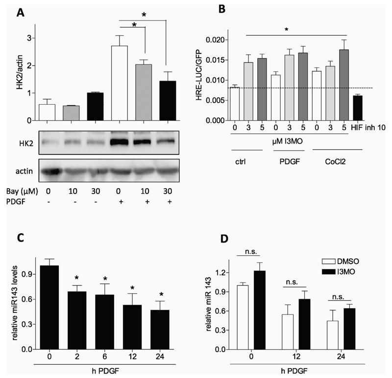
Fig. 6.
HIF1α-and miR143 signaling are not negatively influenced by I3MO. (A) Quiescent VSMC were pretreated with 10 or 30 μM HIF inhibitor BAY 87–2243 and then stimulated with PDGF (10 ng/mL) for 6 h. Total cell lysates were subjected to immunoblot analysis for HK2 and actin. Representative blots and compiled densitometric data from three independent experiments are depicted (n = 3, mean + SD.; * p < 0.05; ANOVA, Bonferroni). (B) CHO cells were transiently transfected with a HRE-LUC reporter gene and EGFP expression construct and serum-deprived. Cells were treated as indicated with I3MO (3 and 5 μM), PDGF (10 ng/mL) and CoCl2 (200 μM) or HIF 1α inhibitor (10 μM) for 8 h before luciferase activity was assessed and corrected for EGFP fluorescence. Bar graph depicts data from three independent experiments. (n = 3, mean + SD.; * p < 0.05; ANOVA, Bonferroni) (C) Quiescent VSMC were treated with PDGF for the indicated periods of time before levels of miRNA 143 were determined as described (n = 5, mean + SD; * p < 0.05; ANOVA, Dunnett vs unstimulated control). (D) Quiescent VSMC were pretreated with DMSO or I3MO for 30 min prior to PDGF stimulation for the indicated periods of time. Then relative miRNA 143 levels were determined as described. (n ≥ 6, mean + SD, n.s. p > 0.05).

