
Figure 2.
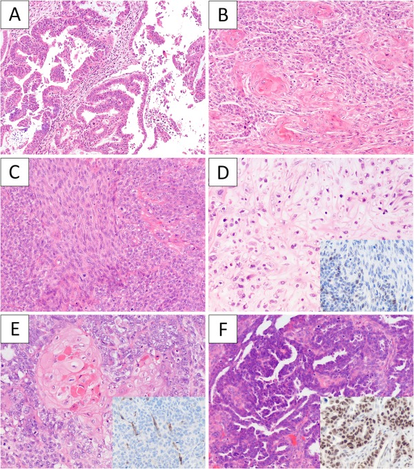
The histological features of selected carcinosarcoma cases. (A) Case 30 showed a low‐grade endometrioid component (A), solid areas with abundant squamous differentiation (B), low‐grade spindle cell component (C) and focal chondroid differentiation (D). p53 immunohistochemistry (D, inset) was focal in both the endometrioid and spindle cell components. Case 22 had uterine, bilateral ovarian and peritoneal tumour. The uterine tumour showed a carcinosarcoma with prototypical endometrioid histology with squamous differentiation (E) and negative staining for WT1 (E, inset). The ovarian tumour as well as the peritoneal tumour deposits were all pure carcinoma with high‐grade serous carcinoma histology (F) and strong nuclear staining for WT1 (F, inset).