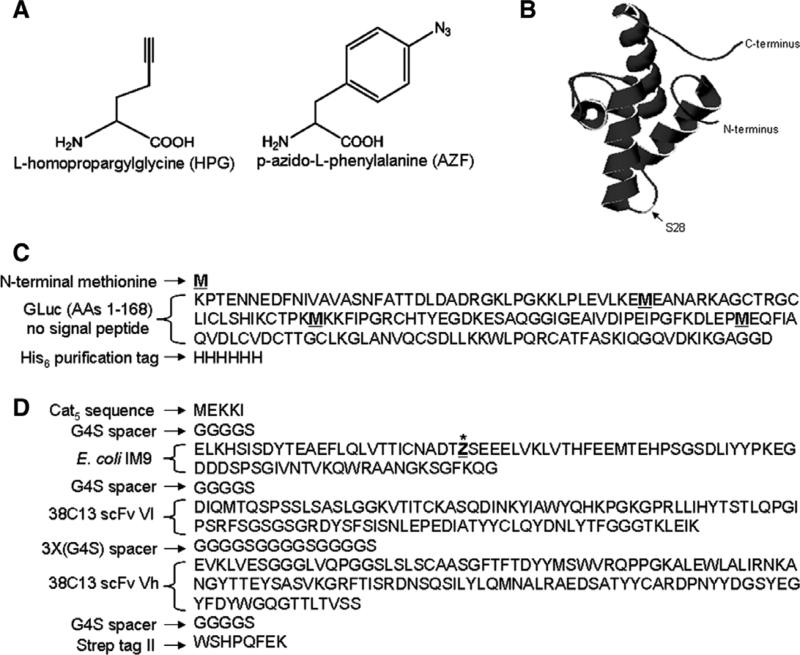
Fig. 1.
(A) Chemical structures of l-homopropargylglycine (HPG) and p-azido-l-phenylalanine (AZF). (B) Crystal structure of E. coli IM9 (PDB ID: 1bxi) showing the surface-exposed serine residue at position 28 (S28). AZF is incorporated in place of S28 in the IM9scFv fusion protein. (C) Amino acid sequence of GLuc. Methionine residues are underlined and indicated in bold. (D) Amino acid sequence of IM9scFv fusion with the site for incorporation of AZF indicated as an underlined “Z” with an asterisk.

