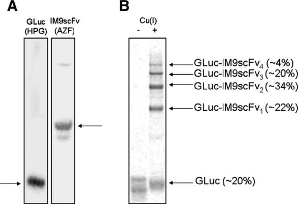
Fig. 3.
(A) Purified GLuc (HPG) and IM9scFv (AZF) proteins, analyzed by SDS–PAGE and coomassie staining. GLuc containing HPG was purified by Ni–NTA chromatography. The IM9scFv fusion protein containing AZF was purified using a Strep-Tactin Sepharose column. Proteins of interest are indicated by horizontal arrows. (B) Autoradiogram of GLuc–IM9scFv conjugates generated by azide–alkyne click chemistry. 3 μM GLuc (HPG) and 24 μM IM9scFv (AZF) samples were incubated with (+) and without (−) Cu(I). The GLuc (HPG) protein was labeled with l-[U-14C]-leucine for visualization by autoradiography. In the presence of Cu(I), the appearance of four bands above the unreacted GLuc suggests that all four HPG residues are accessible for attachment of IM9scFv (AZF). The relative abundance of individual bands is indicated in parentheses.

