Figure 5.

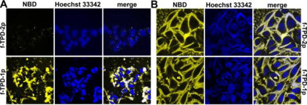
Confocal microscopy images of (A) HepG2 and (B) Saos-2 cells treated with fTPD-2p and fTPD-1p at the concentration of 500 μM for 12 hours. Nuclei are stained by Hoechst 33342.

Confocal microscopy images of (A) HepG2 and (B) Saos-2 cells treated with fTPD-2p and fTPD-1p at the concentration of 500 μM for 12 hours. Nuclei are stained by Hoechst 33342.