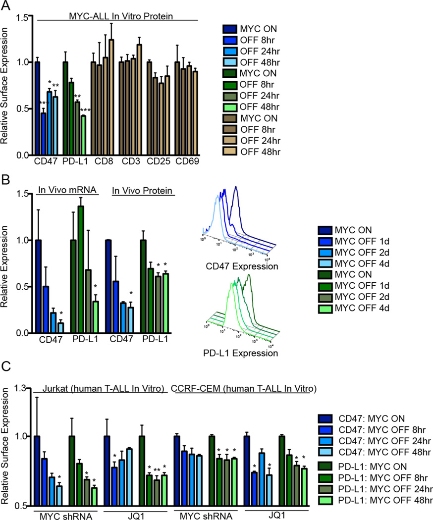
Fig. 1. MYC regulates the expression of CD47 and PD-L1 in murine and human leukemia and lymphomas.
(A) Flow cytometry median fluorescence intensity (MFI) was used to determine the relative cell surface expression of CD47 (blue), PD-L1 (green), and other immune proteins following MYC inactivation in MYC T-ALL 4188 cells in vitro (n=3). (B) Tumors were harvested from primary MYC-driven lymphomas 0 or 4 days following MYC inactivation. mRNA and protein levels were quantified by qPCR and flow cytometry MFI (n=3 tumors per condition). Representative flow cytometry histograms are shown to the right. (C) CD47 (blue) and PD-L1 (green) protein levels in Jurkat and CCRF-CEM cells were quantified by flow cytometry MFI following MYC inhibition by conditional shRNA knockdown or 10 µM JQ1 treatment (n=3 biological replicates).

