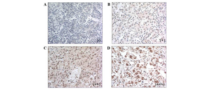
Figure 2.
Immunohistochemical analysis of methyl-CpG binding domain 2 in hepatocellular carcinoma and paratumor liver tissue tissues. Representation of (A) negative staining (−); (B) faintly positive staining (+); (C) moderately positive staining (++); and (D) highly positive staining (+++). Original magnification, ×400.

