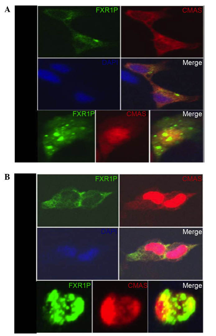
Figure 3.

Colocalization of FXR1P and CMAS in HEK293T and HeLa cells. (A) HEK293T and (B) HeLa cells were transiently transfected with pEGFP-N1-FXR1 and pDsRed-N1-CMAS, then visualized by fluorescence microscopy. FXR1P (green) is visible in the cytoplasm, CMAS (red) is visible mostly in the nucleus, with less in the cytoplasm. Positive colocalization (yellow) is visible in the cytoplasm and around the nucleus. Yellow indicated the colocalization of pEGFP-FXR1P with pDsRed-CMAS. The nuclei (blue) were stained by DAPI. Magnification, ×100. FXR1P, fragile X related 1; CMAS, CMP-N-acetylneuraminic acid synthetase; DAPI, 4′,6-diamidino-2-phenylindole.
