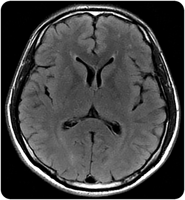
Figure. Representative mild white matter hyperintensities on T2 FLAIR.

Axial T2-weighted fluid-attenuated inversion recovery (FLAIR) MRI brain sequence from a 37-year-old Thai man in Fiebig stage I of acute HIV infection, estimated 14 days postinfection. Punctate white matter T2 hyperintensities in the right and left frontal subcortical white matter were noted. A follow-up MRI brain after 6 months of antiretroviral therapy was unchanged.
