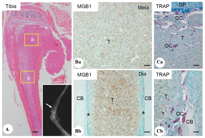
Figure 1.
H&E staining, soft X-ray examination and immunohistochemical analyses of MGB1 and TRAP staining. (A) H&E staining and soft X-ray examination for breast cancer bone metastasis in tibiae. The metastatic tumor tissue occupied the majority of the spaces in the metaphysis and diaphysis. X-ray examination (lower right corner) revealed a radiolucent lesion in the tibia (white arrow). (Ba and Bb) Immunohistochemical analyses for MGB1 in the areas indicated by the yellow boxes in the (Aa) metaphysis and (Ab) diaphysis. Positive MGB1 staining (brown color) indicated metastatic breast cancer tissue. (Ca and b) Histochemical analysis of TRAP. Abundant TRAP-positive osteoclasts were found within the breast cancer metastasis nests and on the surface of the trabecular bone. Scale bars=250 µm for A; 50 µm for B and C. T, tumor cells; CB, cortical bone, *, normal bone marrow tissue; H&E, hematoxylin and eosin; MGB1, mammaglobin 1; TRAP, tartrate-resistant acid phosphatase.

