Figure 2.
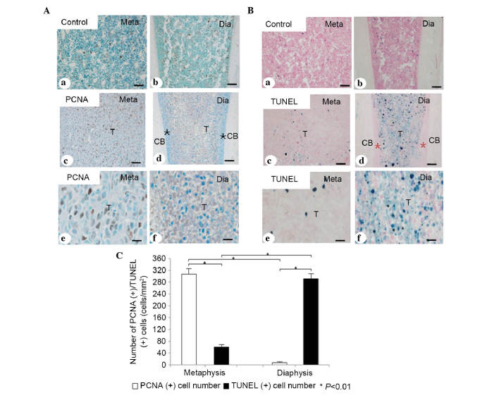
Immunohistochemical and statistical analyses of PCNA and TUNEL staining for apoptosis. (A) Immunohistochemical analysis of PCNA in the (a) metaphysis and (b) diaphysis in the normal bone marrow of the control group. Immunohistochemistry for PCNA in the (c) metaphyseal and (d) diaphyseal tumor metastases. More PCNA-positive tumor cells (brown color) were detected in the metaphyseal tumor tissue, compared with the diaphyseal tumor tissue. (e and f) Higher magnification of c and d, respectively. (B) TUNEL staining for apoptotic tumor cells in the (a) metaphysis and (b) diaphysis in the normal bone marrow tissue of the control group. TUNEL staining for apoptotic tumor cells in the (c) metaphyseal and (d) diaphyseal tumor metastases. More TUNEL-positive apoptotic tumor cells (blue color) were observed in the diaphyseal tumor tissue, compared with the metaphyseal tumor tissue. (e and f) High magnifications of c and d, respectively. (C) Statistical analyses of the numbers of PCNA/TUNEL-positive tumor cells in the metaphysis and diaphysis. *P<0.01. Scale bar=50 µm in Aa-d and Ba-d; 25 µm in Ae and f, and Be and f. TUNEL, TdT-mediated dUTP nick-end labeling; Meta, metaphysis; Dia, diaphysis; T, tumor cells; CB, cortical bone; *, normal bone tissue.

