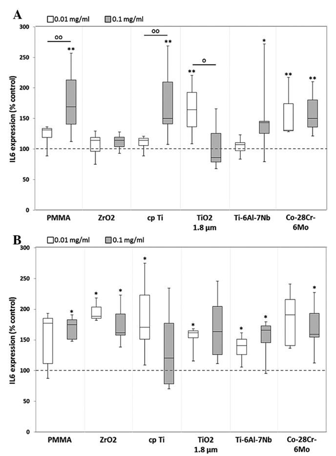
Figure 5.

Release of IL-6 by human (A) osteoblasts and (B) macrophages. Cells were cultivated under standard culture conditions and later treated with the respective particles for 48 h. Subsequently, cell supernatants were collected and analyzed using multiplex enzyme-linked immunosorbent assays (osteoblasts, n≥3; macrophages, n=3). IL-6 contents are displayed relative to the untreated control. Data are presented as box plots. *P≤0.05 and **P<0.01 vs. particle exposure. °P≤0.05 and °°P<0.01 vs. 0.01 mg/ml (Mann-Whitney U test). IL-6, interleukkin-6; PMMA, polymethylmethacrylate; ZrO2, zirconiumoxide; TiO2, titanium dioxide.
