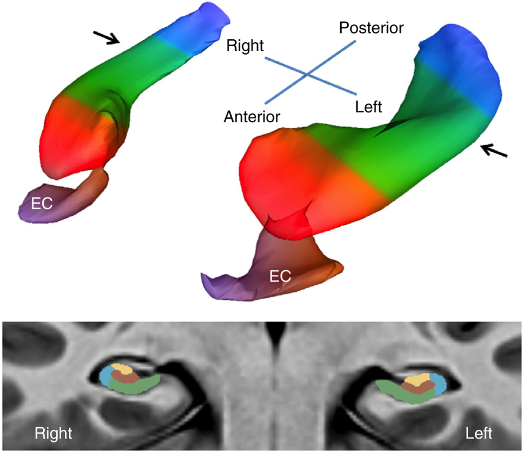
Figure 1.
A bilateral map of the hippocampal circuit generated from the high-resolution acquisitions of CBV-fMRI. A three-dimensional rendering of the bilateral hippocampal circuit (top) derived from a group-wise template of multiple axial slices (illustrated at bottom), generated using the native sub-millimeter resolution of CBV maps (Supplementary Video 1). The EC is the main gateway into the hippocampal circuit, and over the long axis (top) the circuit is divided into the head (red), body (green) and tail (blue). In its transverse axis (bottom), taken through the body of the hippocampal long axis (indicated by arrows, top), the hippocampal circuit is divided into the dentate gyrus (brown), CA3 (yellow), CA1 (blue) and subiculum (green). All statistical analyses were performed only in the boundaries of the hippocampal circuit.

