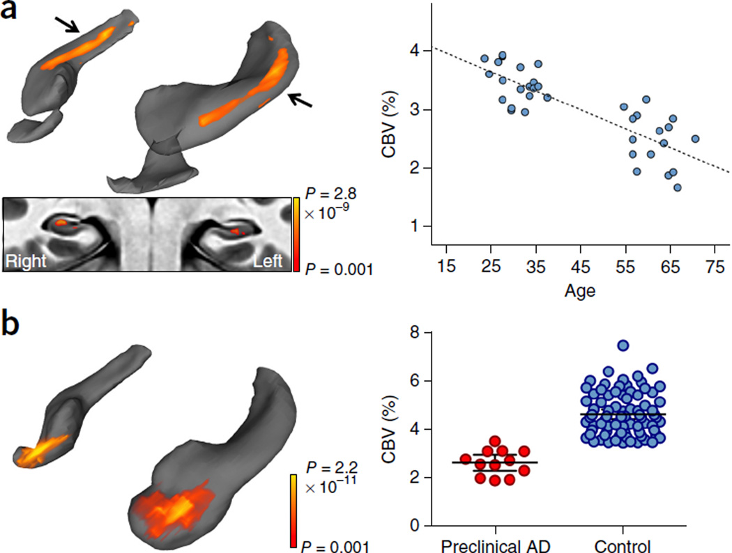
Figure 2.
Mapping a differential pattern of age-related dysfunction in the hippocampal circuit. (a) A voxel-based analysis of 35 individuals ranging from 21–65 years of age revealed that the greatest age-related decline in CBV occurred in the body of the hippocampal circuit (top left; color coded by degree of significance). A transverse slice (bottom left), onto which the hippocampal circuit mask was applied, revealed that age-related CBV decline localized primarily to the dentate gyrus. A scatter plot (right) shows the association between age and mean CBV from all significantly correlated voxels (β = −0.844, r2 = 0.678, P < 0.001). (b) A voxel-based analysis in subjects with preclinical Alzheimer’s disease compared with age-matched controls revealed CBV reductions in the EC (left, reprocessed from ref. 1). A scatter plot (right) shows individual-subject mean CBV values in those lateral EC voxels determined to be significantly different between patients with preclinical Alzheimer’s disease and healthy controls (t94 = 7.265, P < 0.001).

