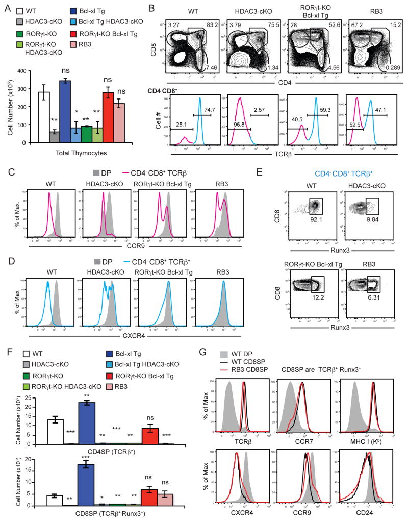
Figure 6. Deletion of RORγt in HDAC3-deficient thymocytes restores thymic cellularity and positive selection of CD8SP thymocytes.
(A) Thymic cellularity of WT, HDAC3-cKO, Bcl-xl Tg, Bcl-xl Tg HDAC3-cKO, RORγt-KO, RORγt-KO HDAC3-cKO, RORγt-KO Bcl-xl Tg, and RB3 (aka RORγt-KO Bcl-xl Tg HDAC3-cKO) mice. Data shown are the mean ± SEM of 4–5 mice per experimental group from four independent experiments. (B) CD4-versus-CD8 profile and TCRβ surface expression of CD4−CD8+ thymocytes from WT, HDAC3-cKO, RORγt-KO-Bcl-xl Tg, and RB3 mice. Data is representative of 4–5 mice per experimental group from four independent experiments. (C) Surface expression of CCR9 on DP and CD4−CD8+ TCRβ− thymocytes from WT, HDAC3-cKO, RORγt-KO, and RB3 mice. Data is representative of at least three mice per group from three independent experiments. (D) Surface expression CXCR4 on DP and CD4−CD8+ TCRβ+ thymocytes from WT, HDAC3-cKO, RORγt-KO, and RB3 mice. Data is representative of at least three mice per group from three independent experiments. (E) Intracellular expression of Runx3 on CD4−CD8+ TCRβ+ thymocytes from WT, HDAC3-cKO, RORγt-KO, and RB3 mice. Data is representative of at least three mice per group from three independent experiments. (F) Quantification of CD4SP (TCRβ+) and CD8SP thymocytes (TCRβ+, Runx3+) from WT, HDAC3-cKO, Bcl-xl Tg, Bcl-xl Tg HDAC3-cKO, RORγt-KO, RORγt-KO HDAC3-cKO, RORγt-KO Bcl-xl Tg, and RB3 mice. Data shown are the mean ± SEM of 4–5 mice per experimental group from four independent experiments. (G) Surface expression of TCRβ, CCR7, MHC class I (Kb), CXCR4, CCR9, and CD24 on DP and CD8SP (TCRβ+, Runx3+) thymocytes from WT mice and CD8SP (TCRβ+, Runx3+) thymocytes from RB3 mice. Data is representation of at least two mice per group.

