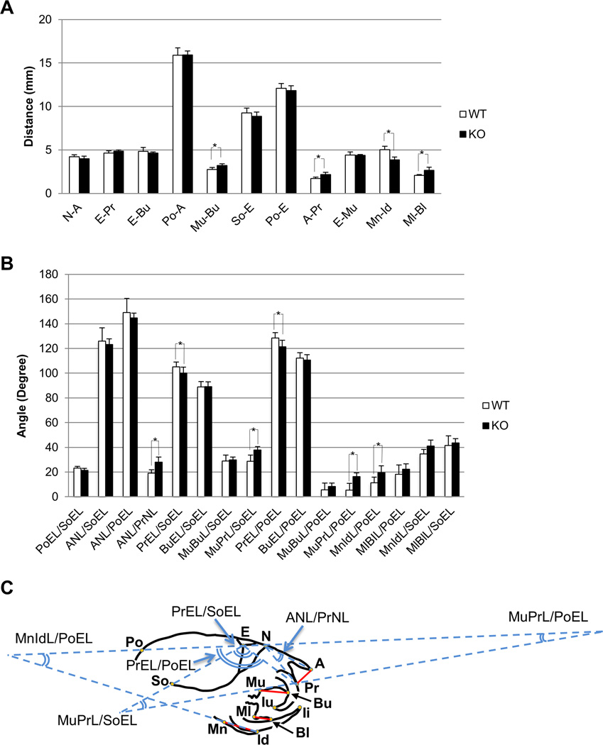
Figure 4. Abnormal Bone-to-bone Relations are not due to Decreased Skull Size in Evc2 KO Mice.
Comparison of linear bone measurements between 1-week-old-WT and 3-week-old-KO mice. (A) No statistical difference was found in the linear bone measurements of N-A, E-Pr, E-Bu, Po-A, So-E, Po-E and E-Mu between WT and KO, while the length of Mu-Bu, A-Pr, and Ml-Bl was significantly higher in KO. * p<0.05. (B) The value of premaxilla relation (PrEL/PoEL) in KO was significantly decreased from that in WT, although the bone measurements in (E-Pr) showed no differences, indicating that the changes in angular bone relation was not entirely due to the bone size. (C) Illustration of the Evc2 KO mouse with reference points and lines in the cephalometric analysis. Red solid lines indicate the linear measurements which were significantly different between 3-week-old-KO and 1-week-old-WT identified in Fig. 4A. Angles in blue lines indicate the angular measurements which were significantly different between 3-week-old-KO and 1-week-old-WT identified in Fig. 4B.

