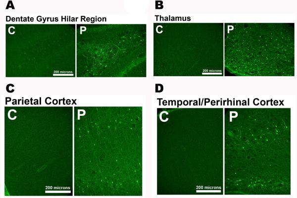
Figure 2.
Neuronal injury following POX-induced SE. Representative photomicrographs of Fluoro-Jade C (FJC) staining in the dentate gyrus–hilus region, parietal cortex, amygdala, and thalamus 2 days after POX SE. Scale bars, 200 μm. Data adapted from Ref. 14.

