
Figure 1. Vascularization micrographs and quantification of time to mid-graft perfusion in long nerve grafts.
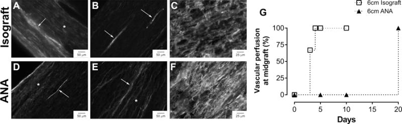
Photomicrographs of longitudinal nerve graft sections after perfusion with EBA and CD31 staining (A–F). Vascular perfusion into the middle of the isograft began by 3 days (A) and was well-established with minimal leakage of EBA by 5 days (B, G). Vascular perfusion was only present in the proximal and distal graft regions of long ANAs at 10 days (D) and completed by 20 days (E, G). Both grafts demonstrated the presence of endothelial cells (CD31) at 5 days regardless of perfusion status (C, F). Arrows indicate perfused, patent vessels, while asterisks indicate leakage of EBA.