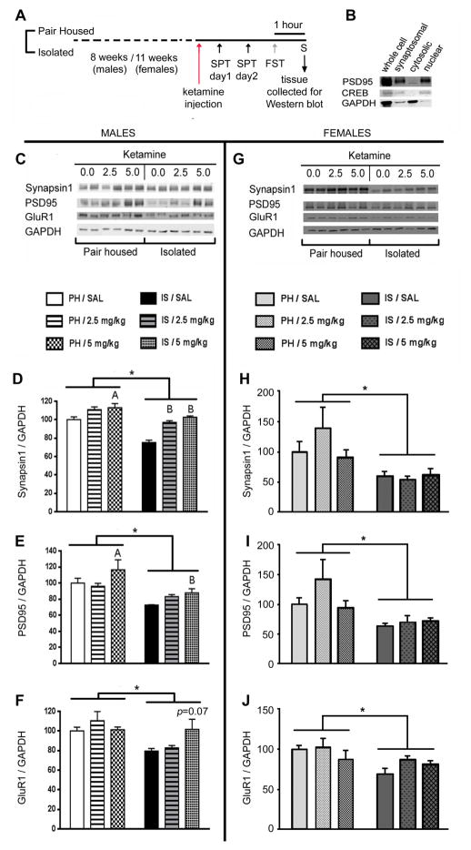
Figure 3.
Ketamine rescued IS evoked decline in synaptic protein levels in males but not in female rats. (A) Schematic representation of the experimental design. Animals were grouped into PH or IS. After 8 weeks (males) or 11 weeks (females) of stress, animals were injected with ketamine (females were injected when they were in diestrus). After completion of behavioral tests(i.e 3 days in males and 3–5 days for females) after ketamine injection, animals were sacrificed and brains were collected for Western blot assays. (B) Synaptoneurosomal fractions collected for Western blot assays were enriched in post synaptic density proteins such as PSD-95. (C) Representative Western blot showing that IS decreases levels of Synapsin1, PSD95 and GluR1 in the mPFC of male rats, and the ability of a single injection of ketamine to reverse this effect. IS evoked a significant decline in levels of (D) Synapsin1, (E) PSD95 and (F) GluR1in male rats; deficits that could be rescued by a single injection of ketamine (5mg/kg). (G) Representative Western blot showing that IS decreases levels of Synapsin1, PSD95 and GluR1 in the mPFC of female rats too, but a single injection of ketamine does not reverse this effect. IS evoked a significant decline in levels of (H) Synapsin1, (I) PSD95 and (J) GluR1 in female rats; however, these deficits could not be rescued by a single injection of ketamine. Levels of individual proteins were quantified using NIH ImageJ. Values represent mean ± SEM (n = 5–7/group) Levels of GAPDH were quantitated to control for differences in amounts of protein loading. (*p<0.05, Ap<0.05 compared to PH/SAL, Bp<0.05 compared to IS/SAL, two-way ANOVA, Fisher’s post-hoc test)

