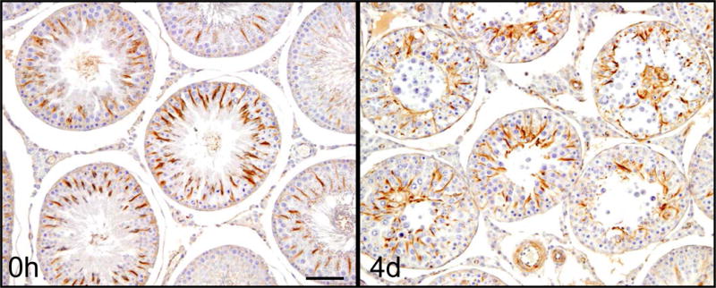
Figure 4. Microtubule disruption after treatment with adjudin.

Rats were administered adjudin, a potential male contraceptive, at a dosage of 50 mg/kg b.w. via oral gavage. Rats were terminated after 4 days, testes removed, fixed, embedded and analyzed by IHC to observe morphological changes in cytoskeleton of seminiferous tubules. The image on the left is a cross section of a normal rat testis stained for α-tubulin. The arrangement of MTs at 0h in normal rat testes shows the typical spoke-like pattern in the Sertoli cell cytoplasm which thus serve as the track for the transport of spermatids and other organelles (e.g., phagosomes, endosomes). MTs lie at an almost 90° angle to the basement membrane of the tunica propria. However, 4 days after adjudin was administered, MT arrangement within the tubules is grossly disrupted, no longer displaying the typical spoke-like pattern. The MT spokes collapsed in concurrence with the loss of spermatids/germ cells, instead of forming columnar structures perpendicular to the basement membrane, MTs had become diffused within the epithelium and many of them are laid in parallel to the basement membrane of the tunica propria. Scale bar, 120 μm, which applies to the other micrograph in this panel.
