Figure 5.
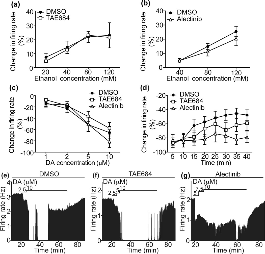
ALK inhibitors attenuate DIR but not the response of DA neurons to acutely administered ethanol or DA. Extracellular recordings of VTA DA neurons were performed in the presence or absence of ALK inhibitors or the DMSO control. (a) Ethanol dose-response curves, showing that ethanol-induced stimulation of DA neuron firing is unaffected by incubation of slices with 100 nM TAE684 (n=10) compared to DMSO controls (n=10). (b) Ethanol dose-response curves, showing that ethanol-induced stimulation of DA neuron firing is unaffected by incubation of slices with 100 nM alectinib (n=11) compared to DMSO controls (n=11). (c) DA dose-response curves, showing that inhibition of DA neurons by DA is unaffected by incubation of slices with TAE684 (open squares, n=9) or alectinib (open triangles, n=8) compared to DMSO controls (solid circles, n=9). (d) Percent change in firing rate over 40 min of exposure to DA in cells treated with DMSO (filled circles, n=9), TAE684 (open squares, n=10), or alectinib (open triangles, n=6). Change in firing rate at 5 min intervals is plotted as a function of time after the initiation of DA administration. DIR was delayed in TAE684-treated and abolished in alectinib-treated slices compared to DMSO-treated controls. (e–g) Representative ratemeter graphs from single neurons showing the effect of DMSO (e), TAE684 (f), or alectinib (g) treatment on DA inhibition. Vertical bars indicate the firing rate over 5 second intervals. Horizontal bars indicate the duration of drug application.

