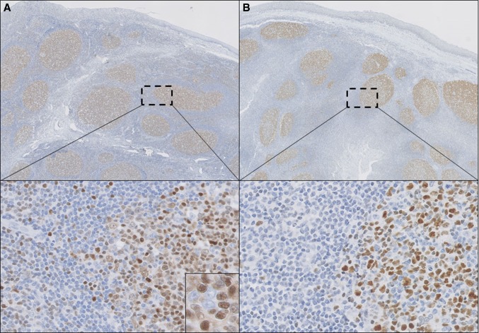
Fig. 1.
Histopathology of a tonsil with reactive follicles. TOX (a) is expressed in reactive follicles, predominantly in centrocytes (inset) in a pattern similar to BCL6 (b), but also in some scattered lymphocytes in the interfollicular areas (selected regions). (a, b ×100, selected regions in a and b ×200 and inset in a ×400)

