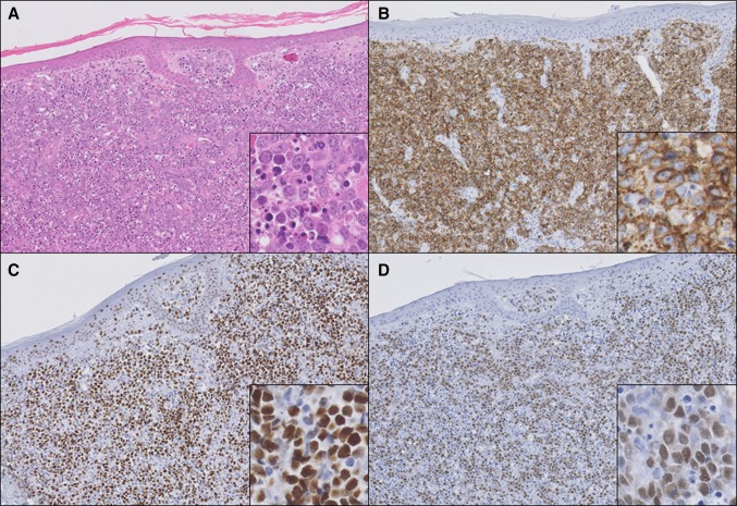
Fig. 3.
Histopathology of a patient with primary cutaneous diffuse large B-cell lymphoma, leg type. Hematoxylin–eosin staining (a) shows diffuse infiltration of the dermis by sheets of CD20+ (b) centroblasts and immunoblasts with many mitotic and apoptotic figures (inset in a). These blastic B-cells stain positive for BCL6 (c) and TOX (d) (a–d ×100, and insets a–d ×400)

