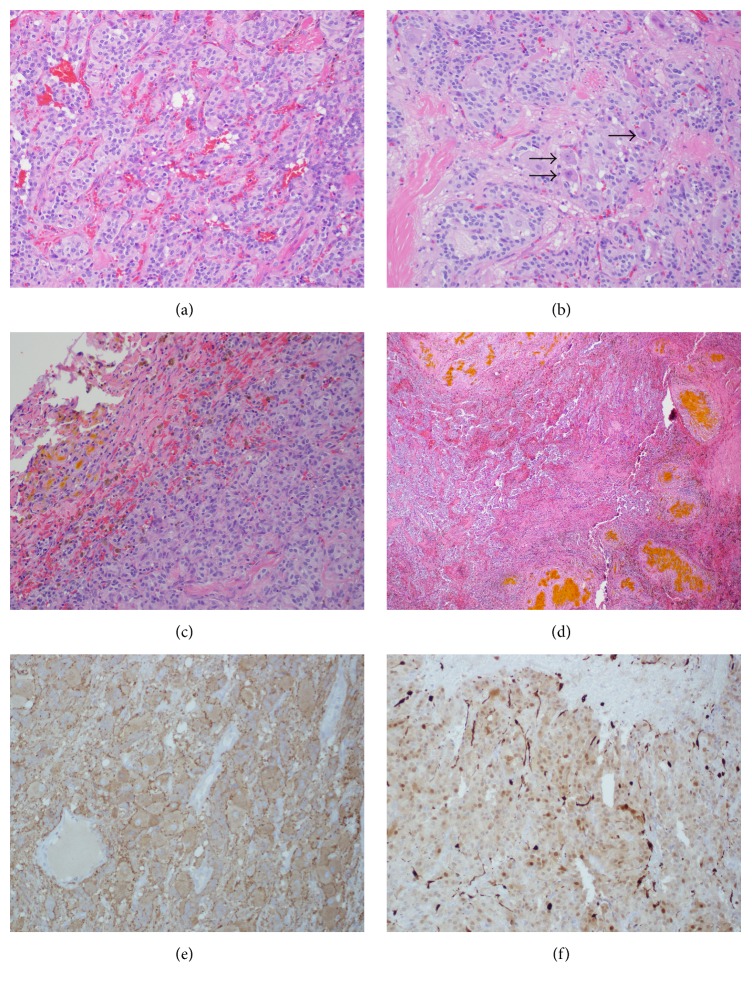
Figure 4.
Gangliocytic paraganglioma. (a) Gangliocytic paraganglioma depicting nested arrangement of cells in a Zellballen pattern. H&E stain, 20x. (b) Gangliocytic paraganglioma with ganglion cells (arrow head). H&E stain, 40x. (c) Section of the tumor capsule showing pigmented macrophages (brown) and extracellular hemoglobin breakdown product (yellow). H&E stain, 4x. (d) Focus of remote hemorrhage toward the periphery of the tumor. H&E stain, 20x. (e) Synaptophysin stain positive (brown): positive for synaptic vesicle protein, 20x. (f) S100 stain positive for sustentacular cells (dark brown along edges of lobules), 20x.

