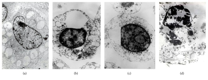
Figure 7.
Transmission electron microscopy images of the Jurkat cells: (a) normal cell morphology; (b) and (c) early apoptosis stages in H2O2-challenged cells representing chromatin condensation and cytoplasmic vacuolization; (d) late apoptosis stage in H2O2-challenged cell representing nuclear fragmentation.

