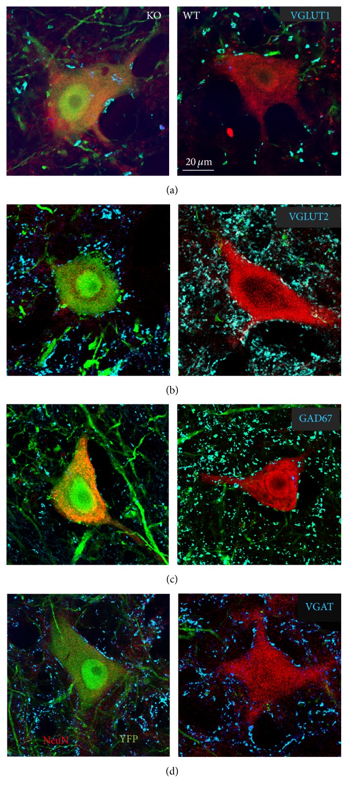
Figure 1.

Immunoreactivity to different synapse-associated antigens surrounding the somata and proximal-most dendrites of large neurons in lamina IX of the L3–5 segments of the spinal cord of SLICK::trkB mice that had been treated with tamoxifen to knock out the trkB gene is shown. Cells expressing YPF (left column) are presumed to be null for the trkB gene and cells in the same histological sections that are YFP− are presumed to have normal expression of this gene. NeuN immunoreactivity (red) is shown to identify these cells as neurons. Because of their large size and laminar location they were considered motoneurons. Synaptic structures immediately adjacent to the perimeters of these cells (cyan) represent contacts made by structures immunoreactive to VGLUT1 (a), VGLUT2 (b), GAD67 (c), or VGAT (d). All cells are shown at the same magnification.
