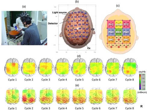
Fig. 1.
Motor learning study using NIRS: (a) experimental setting of the PR, (b) location of optodes, (c) covering cortical surface by each channel, (d) longitudinal cortical activation changes in a healthy subject, and (e) longitudinal cortical activation changes in a stoke patient with ataxia. Reproduced from the articles by Hatakenaka et al.,21,22 where details are reported. PFC, prefrontal cortex; pre-SMA, presupplementary motor area; SMA, supplementary motor area; PMC, premotor cortex; and SMC, sensorimotor cortex.

