
Fig. 1.
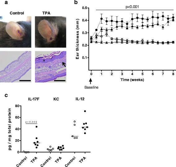
Topical 12-O-tetradecanoylphorbol-13-acetate (TPA) induces local skin inflammation with increased skin thickness and interleukin (IL)-17F levels. a Representative photos illustrating the red and scaly appearance of ears after TPA application as compared to control ears, and representative hematoxylin and eosin-stained ear cross-sections at 10× magnification. Scale bar = 200 μm. TPA led to epidermal hyperproliferation (star) and dermal inflammation (arrow). b Ear thickness (mm) was measured twice weekly in ApoE−/− mice after TPA or vehicle application. Data from two separate, but similar studies with vehicle or TPA application on both ears were included; study 1: n = 5–7/group (unfilled circle: control, filled circle: TPA), study 2: n = 15/group (unfilled triangle: control, filled triangle: TPA). The depicted values in b are mean ± SD, i.e., mean value of right and left ear for each mouse. p < 0.001, control vs. TPA at all time points except at baseline; multiple t-test corrected for multiple comparisons was applied. c Measurement of interleukin IL-17F, IL-12, and keratinocyte-derived cytokine (KC) in ear tissue homogenates after 8 weeks of TPA or vehicle application in study 1. Data is depicted as pg cytokine per mg total protein (median values, unpaired non-parametric t-test)