
Fig. 2.
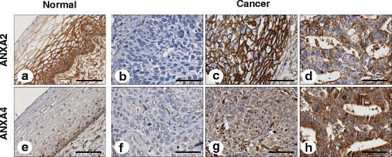
Representative immunohistochemical images of annexin A2 (ANXA2) and annexin A4 (ANXA4) in cervical cancer tissue. ANXA2 expression was strongly detected in the membranes of normal tissues (a), whereas ANXA4 staining was weakly observed in the cytoplasm of normal tissues (e). Negative staining demonstrated a lack of ANXA2 (b) and ANXA4 (f) expression. ANXA2 staining was mainly observed in the membranes of squamous cell carcinoma (c) and adenocarcinoma (d) cervical cancer tissues. ANXA4 staining was restricted to the cytoplasm of squamous cell carcinoma (g) and adenocarcinoma (h) (×200). Scale bar represents 50 μm