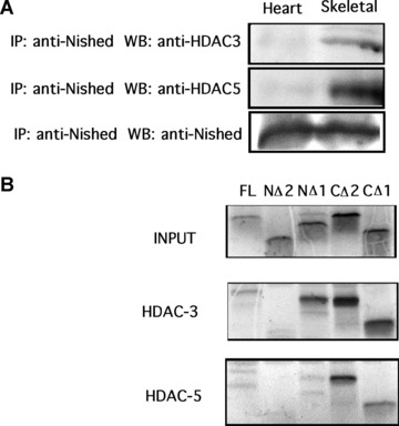
Figure 8(a).

Total cell lystate of mouse cardiac and skeletal muscle tissue was co‐immunoprecipicated with anti‐Nished antibodies and immunoblotted with anti‐HDAC3, anti‐HDAC5 and anti‐Nished antibody (loading control). (B) In vitro transcription/translation pull‐down assay was done using pcNishedV5, CΔ1, CΔ2, NΔ1, NΔ2, pcHDAC3′ flag, pcHDAC5′ flag as described in ‘Material and Methods’. Nished proteins were labelled with S35 methionine and combined with HDAC3 or HDAC5. Proteins were pulled down using M2 anti‐flag agarose, and run on a 18% polyacrylamide gel. Panel 1 (INPUT) shows translated Nished proteins. Panel 2 shows HDAC3 binding to Nished mutants, no binding is seen with NΔ2; panel 3 shows HDAC5 binding to Nished mutant proteins (no binding is seen with NΔ2).
