Figure 5.


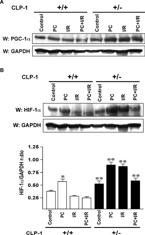
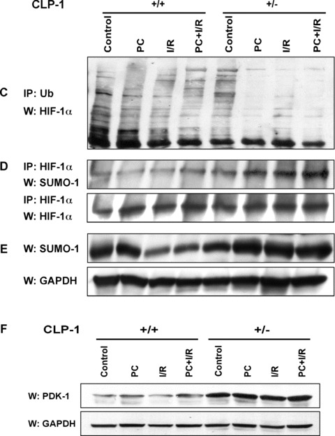
Expression of PGC‐1α and HIF‐1α in heart‐tissue of CLP‐1 heterozygous mice. (A) The expression levels of PGC‐1α in the protein heart extracts from control, preconditioning (PC), ischemia/reperfusion (I/R) and I/R with PC groups of wild‐type and in CLP‐1 +/− hearts was determined by Western blot analysis. GAPDH was used as loading control. (B) We use the same extracts as in Figure 4A to determine the expression levels of HIF‐1α, and as before GAPDH was used as loading control. The experiment was repeated three times and a representative experiment is shown. *P < 0.05 versus wild‐type control, **P < 0.01 versus wild‐type control. (C) The ubiquitination profile of HIF‐1 was examined by using heart extracts from each group. The extracts were used for immunoprecipitation with anti‐ubiquitin antibody followed by Western blot with an antibody against HIF‐1α. (D) The interaction between HIF‐1α and SUMO‐1 was determined by immunoprecipitations using anti− HIF‐1α antibody followed by Western blotting with anti‐SUMO‐1 antibody. Western blot with anti− HIF‐1α antibody served as loading control. (E) Western blot was performed as in (A) to evaluate expression of SUMO‐1. GAPDH was used as loading control. (F) Expression of pyruvate dehydrogenase kinase‐1 (PDK‐1) was evaluated by Western blot using antibody against PDK‐1. The representative figure shows increase in PDK‐1 expression in extracts from CLP‐1 +/− mice hearts subjected to stress. GAPDH expression was used as loading control.
