Figure 3.

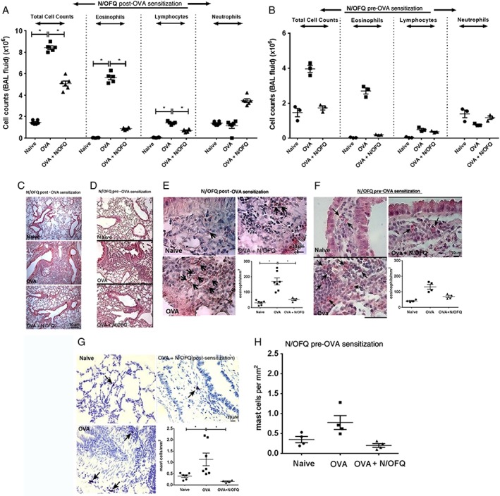
N/OFQ inhibits inflammatory cell infiltration in vivo and recruitment of inflammatory cells within mouse airway tissues. (A) Total and differential cell count in mouse BAL fluid in an in vivo model (N/OFQ post‐OVA sensitization, n = 6 mice), (B) total and differential cell count in mouse BAL fluid in an in vivo model (N/OFQ pre‐OVA sensitization, n = 3 mice). (C) Representative image of HE staining in a N/OFQ post‐OVA sensitized mouse airways (n = 6 mice). Scale bars 50 μm. (D) Representative image of HE staining in a N/OFQ pre‐OVA sensitization mouse airways (n = 3 mice). Scale bars, 50 μm. (E) Modified HE staining showing the kinetics of eosinophilia in a N/OFQ post‐OVA sensitization model (representative image, n = 7 mice) within peribronchial tissue. Scale bars, 10 μ. (F) Modified HE staining showing the kinetics of eosinophilia in a N/OFQ pre‐OVA sensitization model (representative image, n = 4 mice) within peribronchial tissue, scale bars, 10 μm. (G) Representative image of mast cells detected by toluidine blue staining, n = 7 mice. Scale bars, 20 μm. (H) Quantitative estimation of mast cells mm‐2 of mice airway tissue in a N/OFQ pre‐OVA sensitization mouse airways (n = 4 mice). Data expressed as mean ± SEM and analysed by one‐way anova followed by appropriate post hoc tests where relevant. *P <0.05.
