Fig. 1.

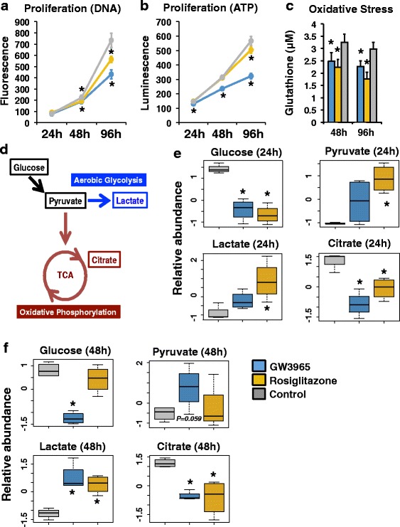
Effects of LXRs and PPARG on cellular phenotypes and metabolites. a Proliferation assays using DNA content after 24, 48, and 96 h of drug treatment (24-h n = 10 for each treatment; 48/96-h n = 12 for each treatment). Luminescence values are presented for GW3965 (blue, LXR agonist), rosiglitazone (gold, PPARG agonist), and DMSO (gray, vehicle control) treatment. *p < 0.01. b Proliferation assays using ATP levels after 24, 48, and 96 h of drug treatment (24-h n = 15 DMSO, n = 8 rosiglitazone, n = 7 GW3965; 48/96-h n = 18 DMSO, n = 9 rosiglitazone, n = 9 GW3965). Luminescence values are presented for GW3965 (blue, LXR agonist), rosiglitazone (gold, PPARG agonist), and DMSO (gray, vehicle control) treatment. *p < 0.01. c Oxidative stress assays after 48 and 96 h of drug treatment (n = 18 DMSO, n = 9 rosiglitazone, n = 9 GW3965). Glutathione concentrations are presented for GW3965 (blue, LXR agonist), rosiglitazone (gold, PPARG agonist), and DMSO (gray, vehicle control) treatment. *p < 0.01. d The carbohydrate metabolism pathway illustrating key metabolites. e Relative metabolite abundance after 24 h of GW3965 (blue, LXR agonist), rosiglitazone (gold, PPARG agonist), and DMSO (gray, vehicle control) treatment. *p < 0.05. f Relative metabolite abundance after 48 h of GW3965 (blue, LXR agonist), rosiglitazone (gold, PPARG agonist), and DMSO (gray, vehicle control) treatment. *p < 0.05. Error bars represent standard deviation
