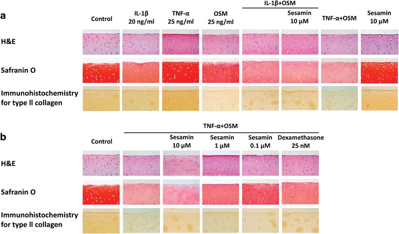
Fig. 6.

Histology staining; H&E staining for chondrocyte cell morphology, Safranin O staining for S-GAGs in cartilage (red color) and immunohistochemistry staining for type ΙΙ collagen remaining in the cartilage (brown color) (x400). a Histology staining of cytokine(s)-induced conditions. b Histology staining of co-treatment of TNF-α and OSM induced-condition with sesamin
