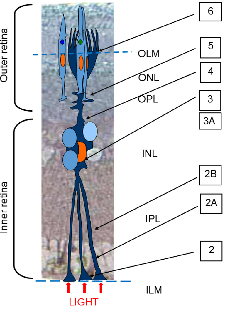
Fig. 1.

Sketch of a Müller cell alignment in the layers of Pied Flycatcher retina; OLM – Outer Limiting Membrane, ONL- outer nuclear layer, OPL- outer plexiform layer, INL- inner nuclear layer, IPL-inner plexiform layer, ILM-inner limiting membrane. Note the locations of the cross-sections of the electron microphotographs presented in the other Figures, marked by the figure numbers.
